
Introduction
At the University of Washington, researchers working on chemical imaging tools for applications like early cancer detection and understanding neurodegenerative disease progression use two-color stimulated Raman scattering (SRS) microscopy. Experiment setups typically include multiple, complex, high-performance instruments for real-time two-color SRS imaging or simultaneous imaging of two widely spaced Raman transitions. Using the Moku:Pro Lock-In Amplifier and Multi-Instrument Mode, they are now able to perform a variety of experiments and extract the low-intensity SRS signals with one compact, multi-channel device.
The challenge
SRS is a coherent Raman scattering process that allows for chemical imaging with both spectral and spatial information. In a typical setup it uses two synchronized pulsed lasers, namely pump and Stokes (Figure 1), to coherently excite the vibration of molecules. To extract the very small SRS signal from a noisy background, a high-frequency modulation and phase-sensitive detection scheme is necessary.
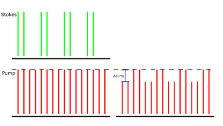
Figure 1: The amplitude modulation transfer of Stokes to the pump beam due to SRS is detected. The demonstrated pump beam has a repetition rate of 80 MHz, and the Stokes beam has the same 80 MHz repetition but is also modulated at 20 MHz. The Δpump is extracted in this detection scheme.
To perform experiments with real-time two-color SRS imaging, researchers must apply orthogonal modulation and detect both in-phase and quadrature signal components.
“In most SRS microscopy experiments, the spectral range is limited to about 300 cm-1 because of limitations in the total bandwidth of the lasers,” said Dr. Dan Fu, Assistant Professor of Chemistry at the University of Washington. “One approach to circumvent this is to scan through wavelengths with a tunable laser, but that is slow and often insufficient for experiments that are time sensitive such as live cell imaging.”
To overcome these limitations, researchers at the University of Washington use a third laser beam to enable simultaneous imaging of two widely spaced spectral regions, for example one in the fingerprint region (e.g. ~1600 cm-1 for Amide vibration) and one in C-H region (e.g. ~2900 cm-1 for protein), but this increases experiment setup footprint and complexity.

Figure 2: HeLa cell SRS images taken with the Moku:Pro Multi-Instrument Mode setup at widely spaced apart Raman transitions.

The solution
A quality lock-in amplifier is a critical hardware component in SRS microscopy experiments with a modulation transfer detection scheme. Moku:Pro’s Lock-in Amplifier provides an intuitive, precise, and robust solution for self-heterodyne signal detection in SRS microscopy experiments. The user interface allows for intuitive and powerful controls for extracting the low-intensity SRS signal.
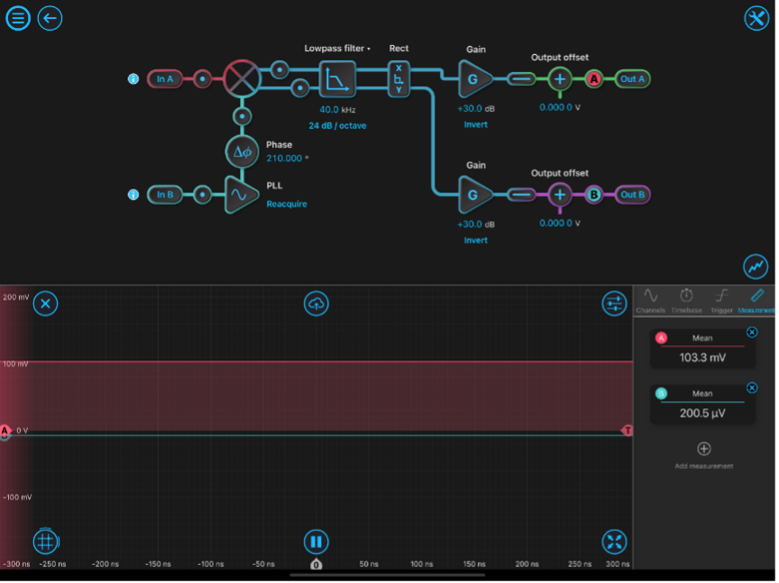
Figure 3: Moku:Pro Lock-in Amplifier with typical single channel configuration settings.
The Moku:Pro Lock-in Amplifier is configured with phase shift, low-pass filter and gain settings optimized for the experiment. The built-in probe points are used for real-time monitoring as setting are adjusted. Both X and Y outputs are made available for dual-channel imaging.
In the case of three lasers, Moku:Pro Multi-Instrument Mode can be configured with two lock-in amplifiers, simplifying the system down to one device without compromises. This allows researchers to take two SRS images of large wave number difference simultaneously, utilizing one Moku:Pro to process two photodiode detector signals.
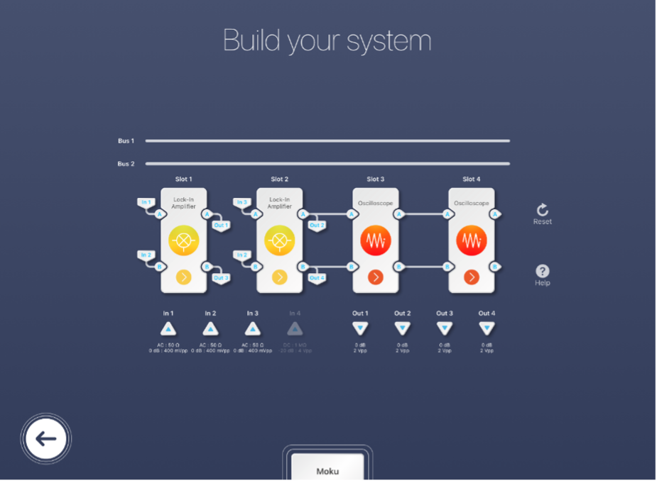
Figure 4: Moku:Pro’s Multi-Instrument Mode with multi-channel lock-in amplifier configuration.
The Multi-Instrument Mode configuration with two lock-in amplifiers for simultaneous SRS microscopy experiments is demonstrated in Figure 4. For the Lock-in Amplifier in Slot 1, input In 1 is the detected signal of the first photodiode, In 2 is the reference, Out 1 is the signal sent to an external data acquisition card, and Out 3 is discarded. For the Lock-in Amplifier in Slot 2, In 3 is the detected signal of the second photodiode, In 2 is once again the reference, Out 2 is the signal sent to external data acquisition card, and Out 4 is discarded. Each detected signal (Out 1 and Out 2) is maximized by adjusting their individual phase shifts before being sent to the data acquisition card. Slot 3 and 4 in this example are configured with Oscilloscopes but can be replaced with another Moku:Pro instrument. 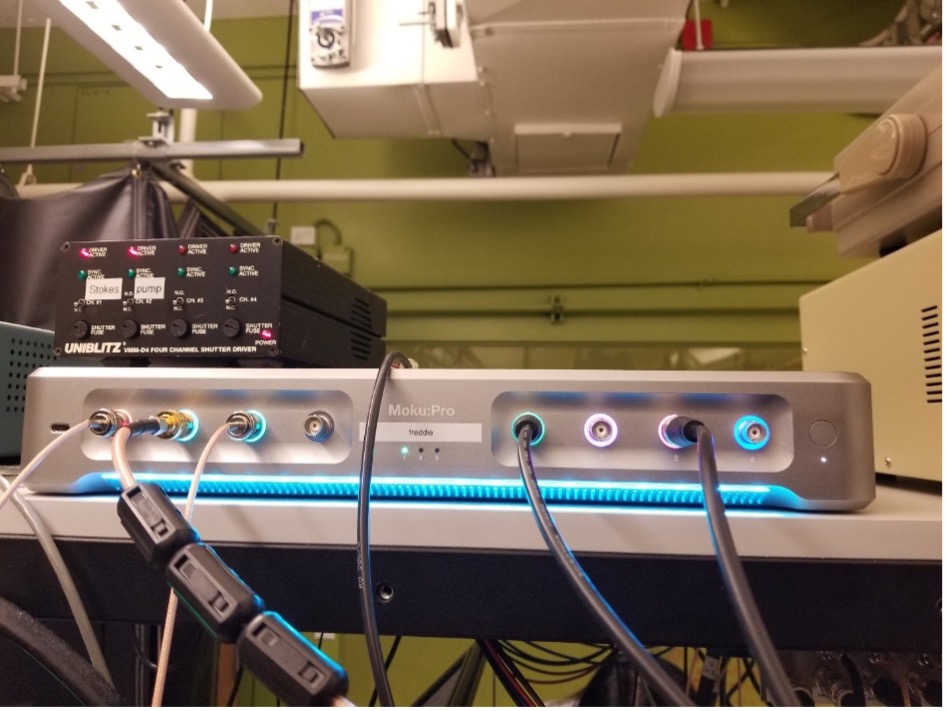
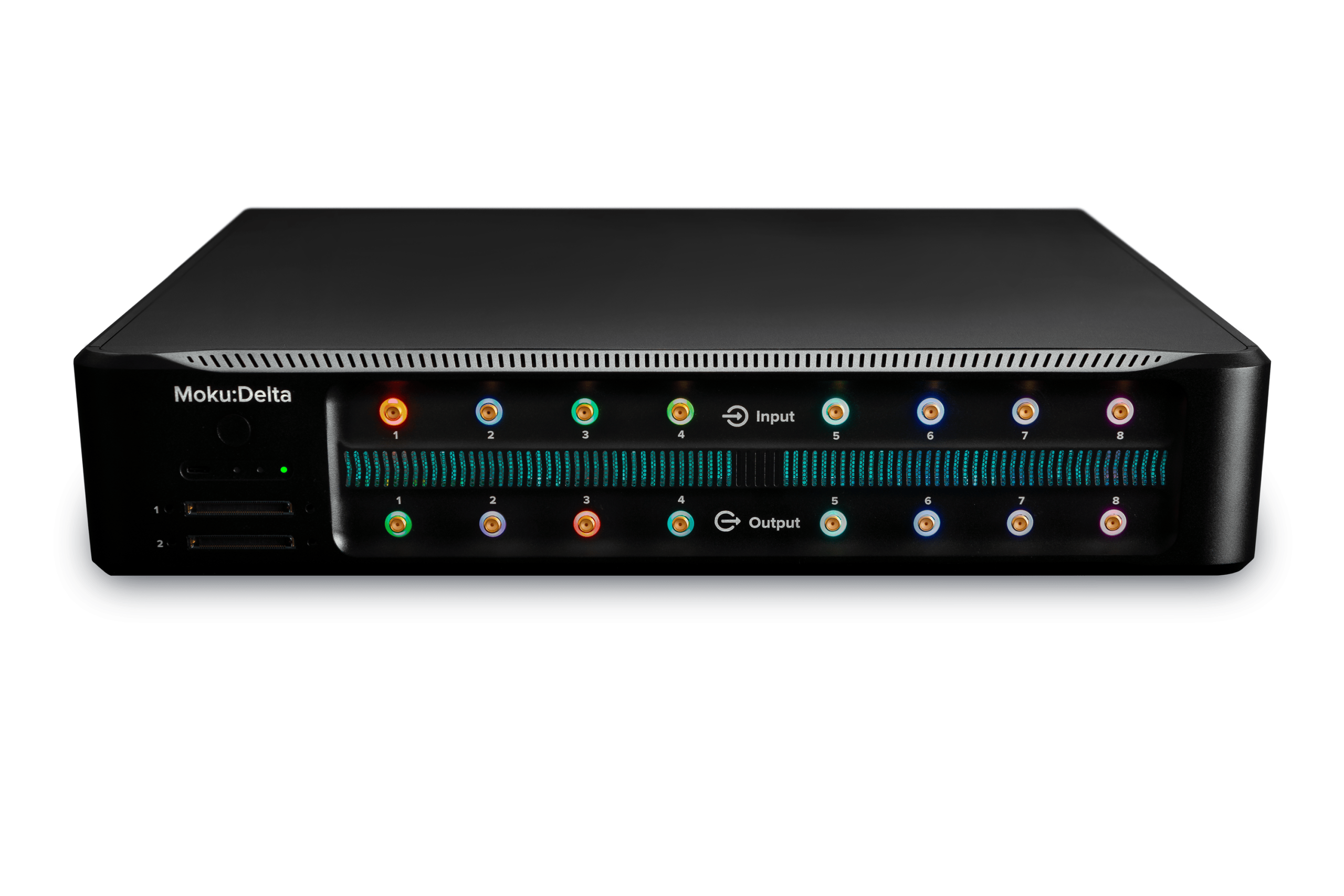
Figure 5: Moku:Pro in Multi-Instrument Mode configured with two lock-in amplifiers with three input channels and two output channels in use.
The result
Moku:Pro’s Lock-In Amplifier provides an excellent solution for a multitude of SRS microscopy experiments. “The user interface allows for intuitive and powerful controls for extracting the low-intensity SRS signal and Moku:Pro’s Multi-Instrument Mode allows for complex imaging experiments on a compact system,” said Dr. Fu. From typical single channel SRS imaging, to dual-channel imaging, and even multi-instrument imaging, researchers at the University of Washington were able to simplify their experiment setup without compromises.